Secondary skin lesions can develop from the primary skin lesions like chronic eczema can lead to lichenification.
The various types of secondary skin lesions are shown in the chart below.

- Skin atrophy: The skin becomes thin and wrinkled. It can occur due to topical steroid use, steroid injection, aging, inflammatory skin conditions, or poor circulation.
- Lichenification: Thickening of the skin with prominent skin markings due to repeated rubbing.
- Excoriation: Straight scratches on the skin that lead to loss of the top layer of skin.
- Crust: A crust forms from the dried exudate, which can be blood, serum, or pus, overlying injured skin, or an irritated skin lesion.
- Erosion: Loss of the top layer of skin called epidermis. The surface is moist and it usually heals without leaving any scar.
- Ulcer: It occurs due to full-thickness loss of the epidermis along with damage to the dermis. It may leave behind scars.
- Fissure: Linear breaks into the skin extending to the dermis (the layer beneath the epidermis). They are usually painful and can occur due to excessive dryness of the skin.
- Scale: Scales appear as white flakes due to increased cell turnover or accumulation of dead skin cells due to abnormal exfoliation.
- Scar: Scars are formed due to the replacement of normal tissue with fibrous tissue. Types of scars include atrophic, hypertrophic, keloid, or contracture.
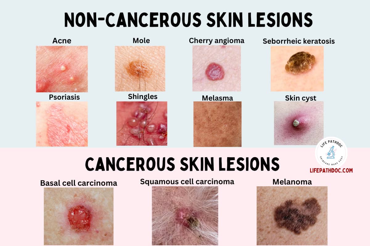
Benign vs Malignant
Benign skin lesions
Benign skin lesions are abnormal skin growths that are non-cancerous. They often grow slowly and are much more common. They do not spread in the body.
Benign skin lesions usually do not require treatment unless they cause symptoms. Some examples include moles, skin tags, lipoma, and cherry angioma.
Malignant skin lesions
Skin lesions that are malignant are skin cancer. They usually grow quickly and the cancerous cells can invade the surrounding tissues and spread in the body.
Malignant skin lesions require treatment. The treatment will depend on the type and stage of the cancer. Some examples include squamous cell carcinoma, basal cell carcinoma, and melanoma.
Diagnosis
Your doctor may diagnose a skin lesion based on the physical examination. They may use instruments like dermatoscope for better examination of the skin lesion.
They also take a complete medical history of your health, any allergies, current medications, or family history.
In some cases, they may do additional testing. These may include:
- Allergy test.
- Culture if an infectious cause is suspected.
- Biopsy of the skin lesion.
Treatment
NEXT PAGE
